 Maple
Maple Peptide degradation pathways fall into three main routes: oxidation of methionine, tryptophan, cysteine and histidine side chains; hydrolytic reactions at aspartate and asparagine residues, which include deamidation, isomerisation and chain cleavage; and physical aggregation driven by hydrophobic and electrostatic interactions. Which route dominates for a given peptide depends on its sequence, the pH and temperature of the solution, and its exposure to oxygen, light and trace metals, so stability has to be assessed compound by compound rather than assumed from a generic shelf life. Understanding these mechanisms is essential for researchers designing experiments that depend on accurate, reproducible peptide concentrations. Maple Research Labs submits manufactured batches of its research peptides to an independent laboratory and publishes the measured result for each tested batch, so that the purity figure a researcher starts from is a measurement rather than a label claim.
Why Peptide Degradation Matters for Research Outcomes
Peptide integrity directly impacts experimental validity. A degraded peptide sample does not simply become “weaker.” Degradation products can include biologically active fragments, inactive species, and potentially immunogenic aggregates that confound results. Researchers working with peptides like BPC-157, GHK-Cu, or semaglutide must account for degradation when interpreting dose-response data, especially in longer-duration protocols.
Zapadka and colleagues (2017, Interface Focus 7:20170030) reviewed the factors that govern the physical stability of peptide therapeutics and identified sequence hydrophobicity, net charge, concentration, pH, ionic strength, temperature and contact with interfaces as the variables that decide whether a peptide stays in solution or aggregates. The same variables apply to research-grade material. In a laboratory the cost of instability is not a failed drug programme but a potency that drifts between experiments and dose-response data that will not replicate.
Pathway 1: Oxidation
Oxidation is the most common chemical degradation pathway for peptides. The amino acids most susceptible to oxidative modification are methionine (forming methionine sulfoxide and, under harsher conditions, methionine sulfone), tryptophan (forming N-formylkynurenine, kynurenine and oxindolylalanine), cysteine (forming disulfide bonds or sulfinic and sulfonic acids), and histidine (forming 2-oxo-histidine).
Methionine Oxidation
Methionine oxidation to methionine sulfoxide is often the first degradation event detected in peptide stability studies. Ji and colleagues (2009, Journal of Pharmaceutical Sciences 98:4485-4500) used parathyroid hormone (1-34) as a model peptide and showed that hydrogen peroxide and tert-butyl hydroperoxide oxidised its two methionine residues preferentially, that peroxide combined with iron extended the damage to tryptophan, and that copper was required before the histidine residue was touched. The most useful part of that work for a research laboratory is its account of where the oxidants come from: degraded polysorbate surfactant, residues of sanitising agents, and metal leached from stainless steel surfaces were identified as realistic sources of peroxide, alkylperoxides and transition metals in a peptide solution. Free methionine added to the solution protected the methionine residues in the model peptide, which is why methionine appears as an excipient in oxidation-prone products. Hovorka and Schöneich (2001, Journal of Pharmaceutical Sciences 90:253-269) reviewed the radical and two-electron mechanisms behind these reactions and the corresponding inhibition strategies: chelation of trace metals, exclusion of oxygen, and sacrificial antioxidants.
Methionine sulfoxide converts a hydrophobic side chain into a polar one. Where the methionine sits in a receptor-binding interface, that change can reduce affinity, but the magnitude is peptide-specific and has to be measured rather than assumed. Peptide chemists have long designed around the problem: the [Nle4, D-Phe7]-alpha-MSH analogue described by Sawyer and colleagues (1980, PNAS 77:5754-5758), the scaffold from which melanotan II was later derived, replaces the methionine of native alpha-MSH with norleucine, an isostere in which a methylene group takes the place of the oxidisable sulfur. For a sequence that retains its methionine, such as thymosin beta-4, the sulfoxide is a defined degradant that a certificate of analysis can resolve; our article on TB-500 methionine oxidation and the sulfoxide degradant covers how it appears on an HPLC trace.
Tryptophan Oxidation
Tryptophan is the residue most susceptible to photo-oxidation. Exposure to UV light or to visible light in the presence of a photosensitiser generates reactive intermediates including N-formylkynurenine and kynurenine. Kerwin and Remmele (2007, Journal of Pharmaceutical Sciences 96:1468-1479) reviewed photodegradation of protein biologics and listed tryptophan, tyrosine, phenylalanine and cysteine/cystine as the primary photo-oxidation targets, noting that photodamage can alter primary, secondary and tertiary structure and that the biopharmaceutical literature on the subject was thin relative to its importance. Pattison, Rahmanto and Davies (2012, Photochemical and Photobiological Sciences 11:38-53) set out the mechanisms in detail: direct photo-excitation of tryptophan and tyrosine, and photosensitised pathways, both radical-mediated and singlet-oxygen-mediated, that damage tryptophan, tyrosine, histidine, methionine and cysteine. The practical point is that light is a controllable input. Amber vials or foil wrapping remove it, and the published mechanism explains why a clear vial left on a bench under fluorescent lighting is a genuine risk for tryptophan-containing peptides such as GHRP-6 or DSIP.
Pathway 2: Hydrolysis
Hydrolytic degradation involves the cleavage or rearrangement of amide bonds by water. While all peptide bonds are theoretically susceptible, certain sequences are dramatically more labile, and the residues that matter most are aspartate and asparagine.
Aspartate-Mediated Cleavage and Isomerisation
The aspartate-proline bond is the classic acid-labile linkage. Piszkiewicz, Landon and Smith (1970, Biochemical and Biophysical Research Communications 40:1173-1178) first documented its anomalous cleavage in dilute acid during protein sequencing, and the reaction is now used deliberately as a chemical fragmentation method. In the pH range that matters for reconstituted peptides, the more important aspartate reaction is not chain cleavage but isomerisation. Oliyai and Borchardt (1993, Pharmaceutical Research 10:95-102) followed the hexapeptide Val-Tyr-Pro-Asp-Gly-Ala across a wide pH range and found that below about pH 3 the dominant reaction was acid-catalysed hydrolysis of the Asp-Gly bond, at pH 4 to 5 hydrolysis and isomerisation through a cyclic imide ran in parallel, and above pH 6 the peptide degraded predominantly by cyclic imide formation leading to the isoaspartyl product. A follow-up study (Oliyai and Borchardt 1994, Pharmaceutical Research 11:751-758) showed that the rate of cyclic imide formation depends mostly on the size of the residue on the carboxyl side of the aspartate, with small residues such as glycine reacting fastest.
Deamidation of asparagine to aspartate and isoaspartate is the other major hydrolytic pathway. Geiger and Clarke (1987, Journal of Biological Chemistry 262:785-794) measured a half-life of only 1.4 days at 37 degrees C and pH 7.4 for the hexapeptide Val-Tyr-Pro-Asn-Gly-Ala, proceeding through a succinimide intermediate that then hydrolysed to a mixture of aspartyl and isoaspartyl products with accompanying racemisation; replacing the glycine after the asparagine with leucine or proline slowed degradation 33- to 50-fold. Robinson and Robinson (2001, PNAS 98:944-949) extended the measurement to 306 asparaginyl sequences in model peptides at pH 7.4 and 37 degrees C and showed that the rate is set primarily by the residue on the carboxyl side, spanning roughly a day for asparagine-glycine to several hundred days for asparagine followed by bulky residues. Because the succinimide can open on either side, the products are isomers with the same mass, which is why deamidation is easy to miss by mass spectrometry alone and is better resolved chromatographically.
Impact on Research Peptides
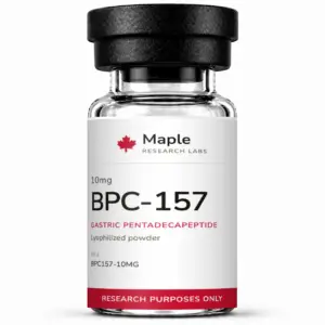
Many research peptides contain asparagine or aspartate residues in their active sequences, and the sequence decides which reaction applies. BPC-157 (Gly-Glu-Pro-Pro-Pro-Gly-Lys-Pro-Ala-Asp-Asp-Ala-Gly-Leu-Val) carries two adjacent aspartate residues and no asparagine, so at physiological pH its relevant slow reaction is aspartate isomerisation to isoaspartate through the cyclic imide, not deamidation, and since the sequence contains no Asp-Pro bond, acid-catalysed chain cleavage is not the primary concern. Researchers running multi-week protocols should factor these kinetics into their experimental timeline, particularly when working with reconstituted solutions at physiological pH, and should not assume that a peptide which still gives the correct mass by MS is chemically unchanged.
Pathway 3: Aggregation
Peptide aggregation is a physical degradation process driven by non-covalent hydrophobic interactions, hydrogen bonding, and electrostatic forces. Unlike oxidation and hydrolysis, aggregation does not alter the primary sequence but changes the peptide’s higher-order state, often irreversibly.
Wang, Nema and Teagarden (2010, International Journal of Pharmaceutics 390:89-99) reviewed the aggregation pathways and the factors that influence them: concentration, temperature, pH, ionic strength, agitation, freeze-thaw stress and contact with surfaces and air-liquid interfaces, with concentration and temperature the most consistently reported accelerants. Zapadka and colleagues (2017) reached the same conclusions for peptides specifically, adding that short peptides with high hydrophobicity and a low net charge near their isoelectric point are the most prone to form fibrils and amorphous aggregates. Aggregation is monitored by size-exclusion chromatography and dynamic light scattering, neither of which is part of a routine purity certificate, so a reversed-phase HPLC purity value says nothing about the aggregation state of a solution.
For research peptides typically reconstituted at microgram-to-milligram-per-millilitre concentrations, the concentration-driven risk is lower than for high-concentration pharmaceutical formulations, but freeze-thaw cycling is a stress that applies at any concentration. Cao and colleagues (2003, Biotechnology and Bioengineering 82:684-690) showed with three model enzymes in dilute solution that the damage depends on the rates involved: slow freezing at about 1 degree C per minute combined with fast thawing above 10 degrees C per minute preserved the most activity, while fast freezing followed by slow thawing caused the most loss, attributed to exposure at the ice-liquid interface and to recrystallisation during thawing. Bhatnagar, Bogner and Pikal (2007, Pharmaceutical Development and Technology 12:505-523) separated the individual stresses of freezing, including cold denaturation, adsorption to the ice surface, freeze-concentration of solutes, and the pH shift that sodium phosphate buffers undergo as one salt crystallises before the other, and reviewed how sugars and surfactants protect against each. Pikal-Cleland and colleagues (2000, Archives of Biochemistry and Biophysics 384:398-406) demonstrated that the freezing-induced pH shift in sodium phosphate buffer was itself sufficient to denature beta-galactosidase, which is a reason to prefer potassium phosphate or non-phosphate buffers for solutions that will be frozen.
Practical Implications: Protecting Research Peptide Integrity
Based on the degradation mechanisms outlined above, researchers can implement several evidence-based storage practices. Store lyophilized peptides at -20 degrees C or below in sealed, desiccated containers to minimize oxidation and hydrolysis; our guide to lyophilization and excipient selection explains why the dried state is so much more stable than solution. Reconstitute in deoxygenated or nitrogen-purged solvent when working with methionine or cysteine-containing peptides. Use amber vials or foil wrapping to protect tryptophan-containing peptides from photo-oxidation. Avoid repeated freeze-thaw cycles by aliquoting reconstituted solutions into single-use volumes, and when a solution must be frozen, freeze it slowly and thaw it quickly. Set the use-by window for a reconstituted solution from stability data for that peptide rather than from a generic number, and in the absence of such data keep refrigerated working solutions short-lived and record the date of reconstitution on the vial. Consider adding 0.1% bovine serum albumin (BSA) as a carrier protein for dilute solutions to reduce surface adsorption losses.
For a detailed guide on reconstitution best practices, see our article on peptide reconstitution: solvent selection and concentration stability.
The Role of COA Verification in Degradation Assessment
A Certificate of Analysis (COA) provides a snapshot of peptide purity at the time of testing, typically by reversed-phase HPLC. However, a COA cannot predict post-purchase degradation. Researchers should interpret HPLC purity values in context: a peptide tested at 99.2% purity that has been stored improperly for 3 months may have degraded significantly by the time it enters an experiment.
This is why Maple Research Labs emphasizes both initial purity verification and proper storage guidance. Manufactured batches are submitted to an independent laboratory, the measured result for each tested batch is published on our certificates of analysis page, and our COA reading guide helps researchers understand exactly what analytical data means for their experimental planning. For researchers new to peptide sourcing, our documentation page provides an overview of our quality standards.
Key Research Findings
Peroxides from degraded polysorbate, residual sanitising agents and metal from stainless steel are the realistic sources of oxidants in a peptide solution, and free methionine protects methionine residues from peroxide (Ji et al., 2009). Tryptophan, tyrosine, phenylalanine and cysteine/cystine are the primary photo-oxidation targets, and light exposure is a controllable variable (Kerwin and Remmele, 2007; Pattison et al., 2012). Asparagine-glycine deamidation proceeds with a half-life of 1.4 days at pH 7.4 and 37 degrees C, and a leucine or proline in place of the glycine slows it 33- to 50-fold (Geiger and Clarke, 1987). Across 306 asparaginyl sequences, the carboxyl-side residue sets the deamidation rate, from about a day to several hundred days (Robinson and Robinson, 2001). Aspartate residues cleave in acid but isomerise to isoaspartate above pH 6 (Oliyai and Borchardt, 1993). Slow freezing with fast thawing preserves the most activity, and the reverse causes the most loss (Cao et al., 2003). The freezing-induced pH shift of sodium phosphate buffer can denature a protein on its own (Pikal-Cleland et al., 2000).
Understanding degradation pathways is not just an academic exercise. It directly informs experimental design, storage protocols, and the interpretation of dose-response data. Maple Research Labs supplies Canadian researchers with starting material whose purity for each tested batch has been measured by an independent laboratory, so that degradation from the source is a documented quantity rather than a confounding variable.
Researchers transitioning from international suppliers can learn more about why Canadian researchers are switching to domestic peptide suppliers for faster delivery and simplified logistics.
Detecting these degradation products depends on the separation principle as much as on the detector. Deamidated and isomerised species that share a retention time with the parent peptide under reversed-phase HPLC are resolved by capillary electrophoresis, which separates by charge-to-size ratio, which is why it serves as an orthogonal purity check. The thermal history that drives the reaction rates above is quantified as mean kinetic temperature across shipping and storage excursions, the least documented interval between certification and receipt.
For research purposes only. Not for human consumption. Not for diagnostic or therapeutic use.
For peer-reviewed research on this topic, visit PubMed.
15% off your first order
Join the research list and we will email your code. New arrivals, current pricing, and research breakdowns. Unsubscribe anytime.
Pingback: Capillary Electrophoresis in Peptide Purity Analysis: Charge-Based Separation, Deamidation Detection, and the Impurities RP-HPLC Cannot Resolve - Maple Research Labs
Pingback: Peptide Photostability: ICH Q1B Light Testing Gaps
Pingback: Vasoactive Intestinal Peptide (VIP) Research | MRL
Pingback: NAD+ and Longevity Peptides | Maple Research Labs
Pingback: Angiotensin-(1-7) Peptide Research: MAS Axis | MRL
Pingback: TB-500 Methionine Oxidation and the Sulfoxide Degradant | Maple Research Labs